




Skip to content
Advertisement
www.nomascom.kz upper
Tribuna.kz

Science & Tech

Chinese researchers develop StableAFM model for atomic force microscopy

StableAFM uses measurement data to recover image detail from fast, sparse scans.

Researchers at the Shanghai Institute of Ceramics of the Chinese Academy of Sciences developed StableAFM for atomic force microscopy. Tests showed that adding physical measurement constraints reduced the model’s mean squared error by about 31.3%.

Source: Chinese Academy of Sciences

Diagram of the StableAFM model for reconstructing atomic force microscopy images
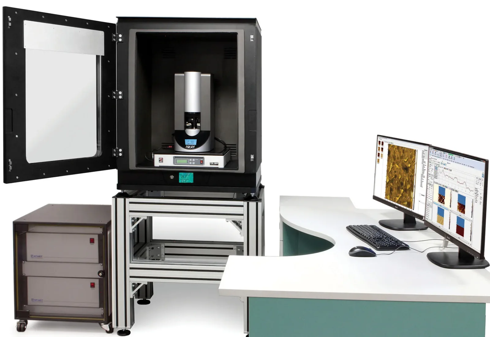
The StableAFM model reconstructs images captured by atomic force microscopy. Illustrative photo. Photo: AAMonitor96 / Wikimedia Commons, CC BY-SA 3.0

Atomic force microscopy (AFM) is used to image nanostructures and characterize material properties, but point-by-point scanning makes it difficult to improve imaging speed and spatial resolution at the same time. A research team at the Shanghai Institute of Ceramics of the Chinese Academy of Sciences has developed StableAFM, a physics-guided generative super-resolution model designed to address this problem.

StableAFM combines AFM sampling physics with a latent diffusion model and uses the original measurement data to constrain image reconstruction. It is designed to recover fine spatial details from fast, sparse scans. The team first uses an AFM-SwinIR network to reconstruct the basic image structure, then applies a diffusion model adapted to AFM images to refine interfaces and local features.

To keep generated details consistent with experimental data, the researchers introduced a latent-space diffusion posterior sampling strategy. It maps each reconstruction back onto the original low-resolution measurement grid and adjusts it according to the difference between the reconstruction and measured data. In tests, the model’s mean squared error fell by about 31.3% when physical measurement constraints were added.


Advertisement
www.toishy.com

The team also used StableAFM to analyze charge traps in the dielectric layers of multilayer ceramic capacitors. It reconstructed high-resolution surface-potential sequences from rapidly collected, low-resolution Kelvin probe force microscopy (KPFM) images. Combined with isothermal surface-potential decay analysis, the sequences revealed the spatial distribution of trap energy levels and density.

The analysis identified a discontinuous, ring-like depletion feature in a cluster of deep-level electron traps near a charge-injection area. The feature appeared in the same region across repeated experiments, while topography images showed no corresponding ring structure. KPFM data collection was four times faster.

The researchers say StableAFM could be applied across materials and imaging modes. It can reconstruct surface scratches and grain boundaries, surface-potential variations in KPFM images, and ferroelectric domain orientation and polarization boundaries in piezoresponse force microscopy (PFM) images. The findings were published in Small Methods.

Translated from Chinese using artificial intelligence. Source: Chinese Academy of Sciences

Countries:

Read also

Advertisement
Nomascom banner footer